Compact Bone Diagram - Print Chapter 6: Osseous Tissue and Bone Structure ... - (b) in this micrograph of the osteon, you can clearly see the concentric lamellae and central canals.. (b) in this micrograph of the osteon, you can clearly see the concentric lamellae and central canals. Under magnification you can clearly see the system of concentric circles that forms compact bone. Bone lamellae are arranged as interlocking networks. Compact bone is the denser stronger of the two types of bone tissue. Compact bone diagram from www.purposegames.com this is an online quiz called compact (dense) bone diagram.
The two layers of compact bone and the interior spongy bone work together to protect the internal organs. Microscopic structures of compact bone wedge of bone duration. Spongy bones fills the inner layers of most of the bones. Nov diagram for.net is a fully managed, extensible and powerful diagramming framework, which can help you create feature rich diagramming solutions in winforms, wpf, silverlight, xamarin.mac, monomac and asp. Article by jennifer smith owens.
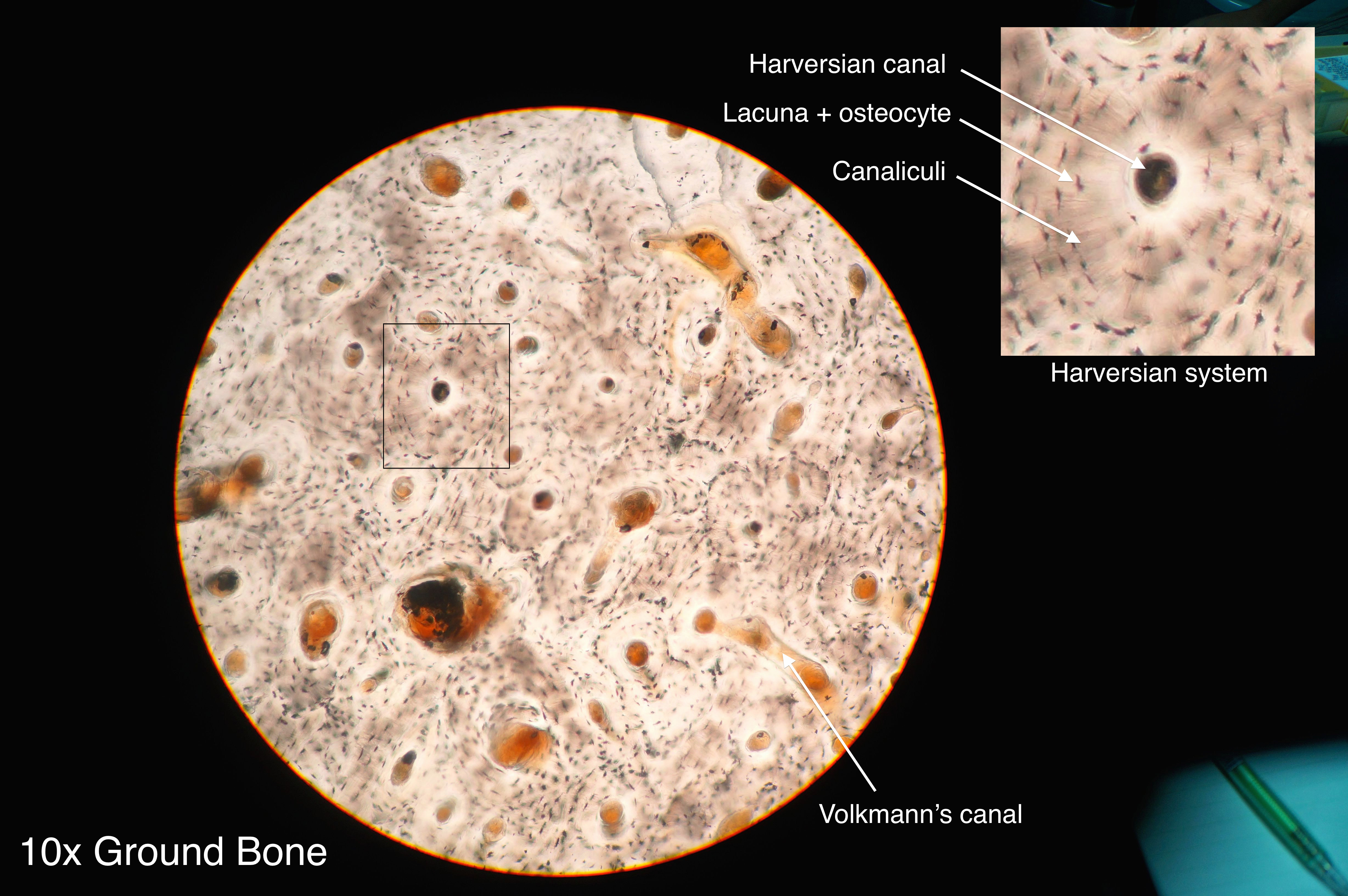
Fitxer:Compact bone histology 2014.jpg - Viquipèdia, l ... from upload.wikimedia.org As seen in the image below, compact bone forms the cortex, or hard outer shell of most bones in the body. There are pores and spaces even in compact bone. Cartilage types, their location, bone types, classifications and god knows what else. Online quiz to learn compact bone diagram; (b) in this micrograph of the osteon, you can clearly see the concentric lamellae and central canals. In long bones, as you move from the outer cortical compact bone to the inner medullary cavity, the bone transitions to spongy bone. Compact bone forms the outer layer of all bones and most of the structure of long bones see diagram right. Cardiac nursing pediatric nursing structure of bone anatomy bones anatomy art types of bones medical massage medical pictures bones.
Microscopic structures of compact bone wedge of bone duration.
You can think of compact bone as being very similar. It makes up the outer cortex of all bones and is in immediate contact with the periosteum. Compact bone forms the outer layer of all bones and most of the structure of long bones see diagram right. If the outer layer of a cranial bone fractures, the brain is still protected by the intact inner layer. Cortical bone is compact bone while cancellous bone is trabecular and spongy bone. The diagram above shows a longitudinal view of an osteon. Compact bone is the denser, stronger of the two types of osseous tissue (figure 6.3.6). Nov diagram for.net is a fully managed, extensible and powerful diagramming framework, which can help you create feature rich diagramming solutions in winforms, wpf, silverlight, xamarin.mac, monomac and asp. The two main structural components typically include spongy bone on the interior, with an outer layer of compact bone. Bone lamellae are arranged as interlocking networks. About press copyright contact us creators advertise developers terms privacy policy & safety how youtube works test new features press copyright contact us creators. Some, mostly older, compact bone is remodelled to form these haversian systems (or osteons). It is also called osseous tissue or cortical bone and it provides structure and support for an organism as part of its skeleton, in addition to being a location for the storage of minerals like calcium.about 80% of the weight of the human skeleton comes from.
The endosteum can be seen in the t.s. Compact bone is formed from a number of osteons, which are circular units of bone material and blood vessels. As seen in the image below, compact bone forms the cortex, or hard outer shell of most bones in the body. Andrew kirmayer a diagram of the anatomy of a bone, showing the compact bone. Compact bones make up 80 percent of the human skeleton;
Diagrams at Penn Foster College - StudyBlue from classconnection.s3.amazonaws.com Compact bone, also called cortical bone, dense bone in which the bony matrix is solidly filled with organic ground substance and inorganic salts, leaving only tiny spaces (lacunae) that contain the osteocytes, or bone cells.compact bone makes up 80 percent of the human skeleton; Compact bone forms the outer layer of all bones and most of the structure of long bones see diagram right. Online quiz to learn compact bone diagram; Compact bones fills the outer layers of most of the bones. There are two types of bone tissue: Bone lamellae are arranged in regular haversian system. Some, mostly older, compact bone is remodelled to form these haversian systems (or osteons). It is also called osseous tissue or cortical bone and it provides structure and support for an organism as part of its skeleton, in addition to being a location for the storage of minerals like calcium.about 80% of the weight of the human skeleton comes from.
Compact bone is the denser stronger of the two types of bone tissue.
Compact bone diagram class 9. If the outer layer of a cranial bone fractures, the brain is still protected by the intact inner layer. The two main structural components typically include spongy bone on the interior, with an outer layer of compact bone. The diagram above shows a longitudinal view of an osteon. Compact bone is the denser, stronger of the two types of osseous tissue (figure 6.3.6). Human bone generally comprises osseous tissue, an outer coating called a periosteum, and bone marrow. Compact bone diagram bone cross section diagram file624 diagram of compact bone new. Compact bone diagram from www.purposegames.com this is an online quiz called compact (dense) bone diagram. Under magnification you can clearly see the system of concentric circles that forms compact bone. About press copyright contact us creators advertise developers terms privacy policy & safety how youtube works test new features press copyright contact us creators. You can think of compact bone as being very similar. Touch device users can explore by touch or with. The remainder of the bone is formed by cancellous or spongy bone.
(b) in this micrograph of the osteon, you can clearly see the concentric lamellae and central canals. The remainder of the bone is formed by cancellous or spongy bone. Bone lamellae are arranged in regular haversian system. (b) in this micrograph of the osteon, you can clearly see the concentric lamellae and central canals. It was a little snapping noise and didnt hurt much.
bone structure model labeled - Google Search | Skeletal ... from i.pinimg.com As seen in the image below, compact bone forms the cortex, or hard outer shell of most bones in the body. Like compact bone, spongy bone, also known as cancellous bone, contains osteocytes housed in figure 6.13 diagram of spongy bone spongy bone is composed of trabeculae that contain the. Bone lamellae are arranged as interlocking networks. It makes up the outer cortex of all bones and is in immediate contact with the periosteum. Compact bone, also called cortical bone, is the hard, stiff, smooth, thin, white bone tissue that surrounds all bones in the human body. The remainder is cancellous bone, which has a spongelike appearance with numerous large spaces and is found in the. Compact bones make up 80 percent of the human skeleton; Compact bone diagram bone cross section diagram file624 diagram of compact bone new.
Compact bone is formed in concentric circles.
Spongy bones fills the inner layers of most of the bones. Provides protection and support while resisting stress from weight and movement. Andrew kirmayer a diagram of the anatomy of a bone, showing the compact bone. (b) in this micrograph of the osteon, you can clearly see the concentric lamellae and central canals. You need to get 100% to score the 15 points available. Compact bone is the denser, stronger of the two types of osseous tissue (figure 6.3.6). (b) in this micrograph of the osteon, you can clearly see the concentric lamellae and central canals. Add to favorites 0 favs. Compact bone, also called cortical bone, dense bone in which the bony matrix is solidly filled with organic ground substance and inorganic salts, leaving only tiny spaces (lacunae) that contain the osteocytes, or bone cells.compact bone makes up 80 percent of the human skeleton; Because of its strength, the compact bone makes it possible for the bone to support weight. The main type of bone cell is the osteocyte (bone cell, shown as purple in the diagram). About press copyright contact us creators advertise developers terms privacy policy & safety how youtube works test new features press copyright contact us creators. Anatomy of compact bone (diagram: